Donar
DonarLaboratorio de anatomía vegetal del Jardín Botánico de Santa Bárbara
En una pequeña y discreta sala del lado oeste del Jardín hay un minúsculo laboratorio que ha influido enormemente en la forma en que los científicos ven el mundo. ¿Es una exageración? En realidad no: durante más de 25 años, el Laboratorio de Anatomía Vegetal del Jardín ha sido la base del trabajo anatómico pionero del Dr. Sherwin Carlquist (1930-2021), investigador asociado, y de otros investigadores. Sherwin se instaló en el Jardín tras su jubilación de la Universidad de Claremont en 1993. Desde entonces hasta su muerte a los 91 años, Sherwin y sus colegas publicaron más de 179 artículos utilizando datos generados en su mayor parte en este humilde espacio. La necrológica de Sherwin en el diario Independent describe magníficamente sus contribuciones a la ciencia, el arte y la humanidad.
¿Qué hay en el Laboratorio de Anatomía Vegetal?
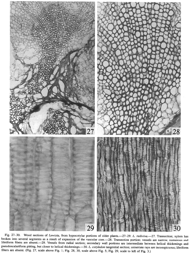
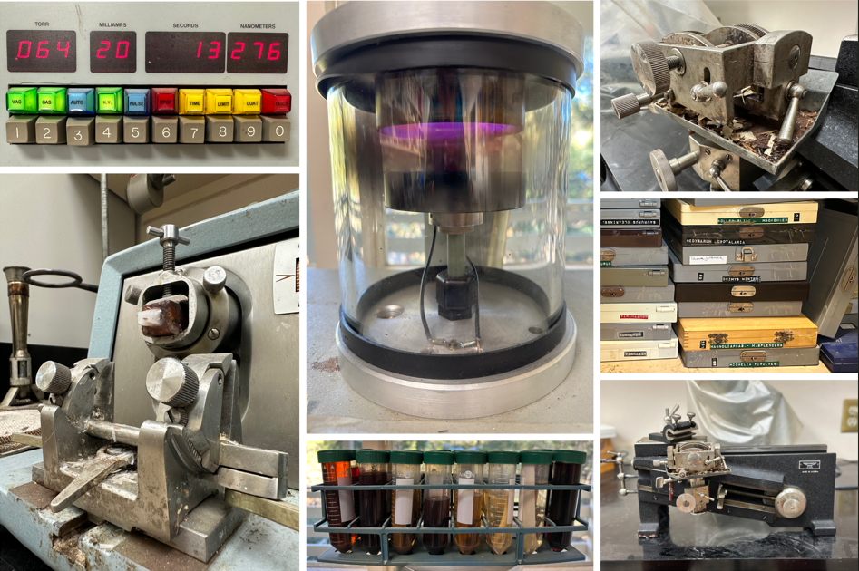
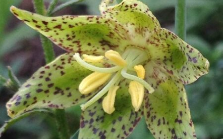
Esta pequeña sala contiene casi todas las herramientas que necesita un botánico para explorar el funcionamiento microscópico interno de una planta. Sherwin se centró principalmente en la vasculatura de las plantas: los diminutos tubos que transportan agua y minerales desde las raíces hasta los brotes (el xilema), así como los que llevan los azúcares producidos en las hojas mediante fotosíntesis al resto de la planta (el floema) (Foto Panel 1). Aunque uno de los principales objetivos de Sherwin era describir y contextualizar esta "fontanería vegetal" a través de la biodiversidad vegetal mundial, el planteamiento funciona igual de bien para investigar otras partes de la planta (por ejemplo, las flores). Los aparatos anatómicos estándar son deliciosamente steampunk: manivelas accionadas a mano; muestras de madera sujetas con abrazaderas que se deslizan sobre bastidores enrojecidos por el aceite para cortar finos trozos de madera con cuchillas afiladas a mano; bandejas metálicas para calentar portaobjetos de la época de 1950. En el panel fotográfico 2 se muestran algunas imágenes de estas herramientas de trabajo.
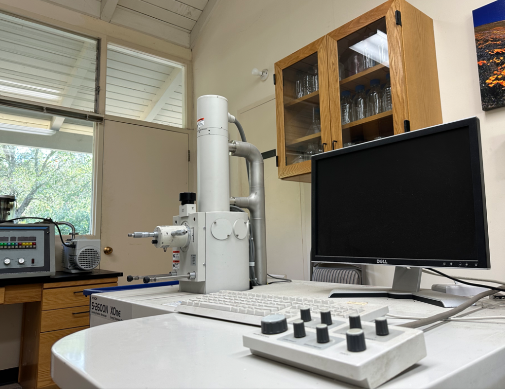
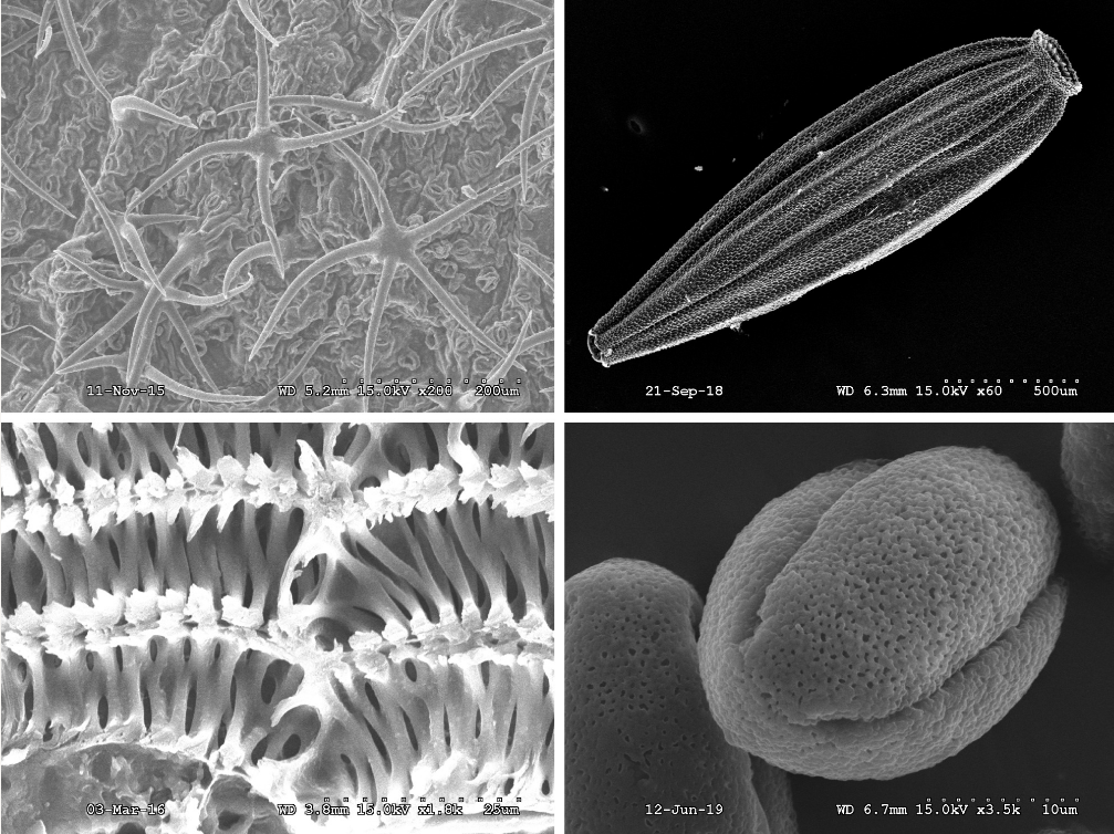
Junto a estos pilares del laboratorio de anatomía se encuentra el microscopio electrónico de barrido (SEM; Foto 3). La microscopía óptica tradicional es ideal para explorar objetos algo más grandes (pequeños), pero el MEB toma el relevo para los verdaderamente diminutos, donde pueden ser necesarios aumentos de 5.000 a 10.000 veces o más. El MEB enfoca un haz estrecho de electrones sobre una muestra recubierta de oro colocada al vacío, con sensores que interpretan la dispersión de electrones para revelar la superficie de la muestra con exquisito detalle. Aquí hay un vídeo rápido de YouTube que explica cómo funciona un SEM. El panel fotográfico 4 muestra algunas imágenes generadas por científicos del Jardín utilizando nuestro SEM.
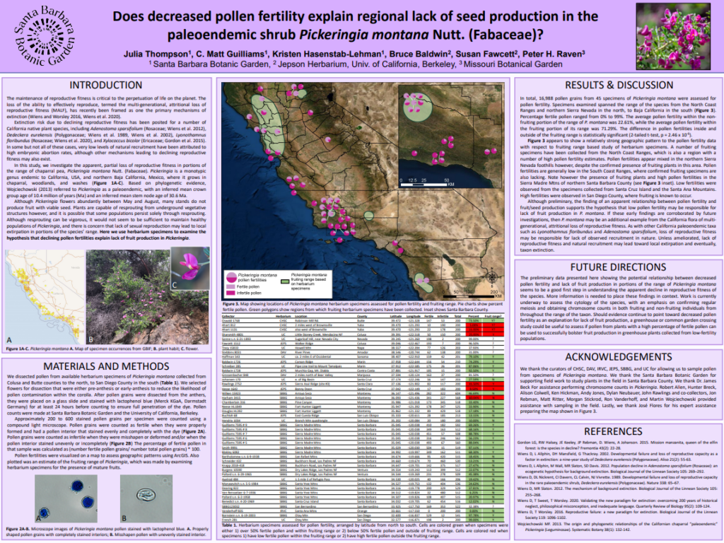
Las herramientas y técnicas empleadas en el Laboratorio de Anatomía Vegetal son tan pertinentes hoy como cuando Sherwin reutilizó el espacio a mediados de los años noventa. Los científicos del Departamento de Conservación e Investigación siguen utilizando este laboratorio para investigar muchos aspectos de la morfología y la anatomía de las plantas. Algunos estudios recientes ponen de relieve la importancia de este espacio para nuestro trabajo. Durante el verano de 2022, la becaria de Conservación de Jardines Julia Thompson, guiada por el Equipo de Biodiversidad y colaboradores, investigó la causa del aparente fracaso reproductivo del guisante chaparral (Pickeringia montana), que ya no produce semillas en algunas partes de su área de distribución. Julia utilizó el laboratorio de anatomía para evaluar la fertilidad del polen del guisante de los chaparrales, corroborando observaciones preliminares anteriores de que las poblaciones que no producen frutos también tienden a tener una fertilidad reducida del polen. Con una mejor comprensión de la causa del fracaso reproductivo en la mano, los botánicos pueden ahora ser capaces de diseñar una intervención para prevenir la extirpación local de la especie. Julia presentó nuestros hallazgos en el Simposio de Botánicos del Sur de California de 2022, donde su presentación ganó el primer puesto en el concurso de carteles. La foto 5 muestra nuestro póster.
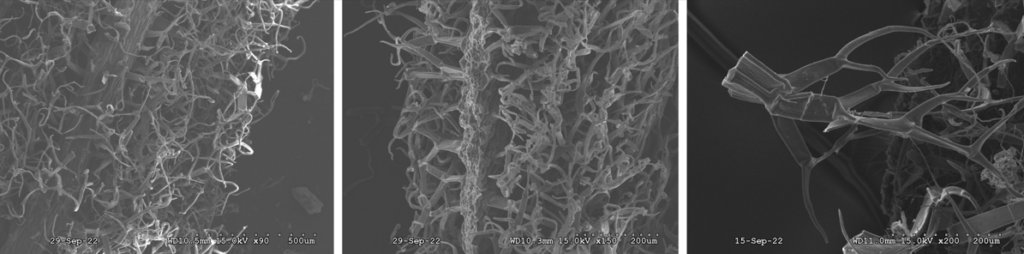
Caitlin Hazelquist, técnica del Laboratorio de Genética de la Conservación, utiliza el microscopio electrónico de barrido en el Laboratorio de Anatomía Vegetal para estudiar las características microscópicas de las hojas de la planta endémica de la isla de Santa Rosa en peligro de extinción, el pincel de hojas blandas (Castilleja mollis). Como puede sugerir su nombre común, esta especie tiene hojas recubiertas por una capa aterciopelada de pelos intrincadamente ramificados, una posible adaptación al entorno marítimo en el que crece. El MEB nos ofrece una visión sin precedentes de la arquitectura ramificada de los pelos, que podría ayudarnos a diferenciar los híbridos con una especie afín de pincel. Dado que la hibridación entre el pincel de hojas blandas y sus parientes cercanos puede estar acelerándose con el cambio climático, es fundamental identificar claramente las plantas híbridas sobre el terreno utilizando las características de las hojas. La lámina 6 muestra algunas de las magníficas imágenes SEM de Caitlin.
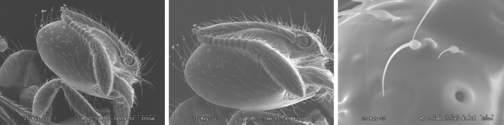
El equipo de ecología también utiliza el laboratorio para visualizar estructuras microscópicas con el microscopio electrónico de barrido y para utilizar la campana extractora de humos químicos del laboratorio para secar insectos de forma segura. El ecólogo especialista en conservación de invertebrados terrestres Zach Phillips prevé utilizar el SEM para obtener imágenes de las diminutas características de algunos invertebrados que estudia, como las hormigas del condado de Santa Bárbara. El panel fotográfico 7 muestra algunas de las imágenes SEM recientes de Zach, incluida una imagen a 1800' de aumento de uno de los espiráculos de las hormigas, que son pequeñas aberturas en el tórax y el abdomen utilizadas para respirar. Un material inspirador.
Para más información sobre el trabajo del Jardín en el Laboratorio de Anatomía Vegetal, póngase en contacto con el Sistemático de Plantas de Tucker y Conservador del Herbario, Matt Guilliams. | mguilliams@sbbotanicgarden.org.
Apoyo a los polinizadores y a la fauna silvestre local mediante plantas autóctonas
No todas las plantas arden igual: una mirada a nuestra investigación sobre seguridad contra incendios
Donante destacado: Devolver a los lugares que importan